Leukemia Blood Cells Detection using Neural Network Classifier
DOI:
https://doi.org/10.37934/araset.33.1.152162Keywords:
Leukemia, blood cells, neural networkAbstract
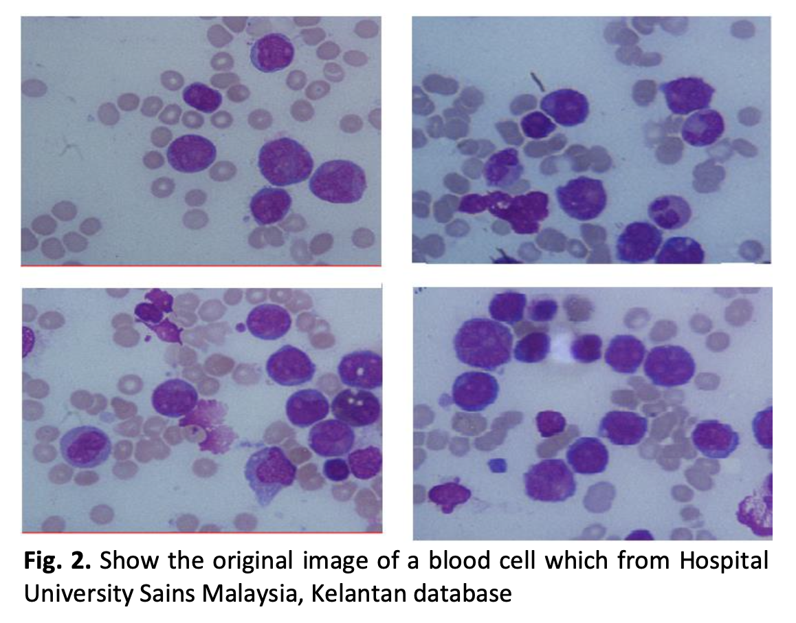
Image segmentation is an image processing operation performed on the image in order to partition the image into some images based on the information contained in the original image. Image segmentation plays an important role in many medical imaging applications, image segmentation facilitates the anatomy process in a particular body of human body. Classification and clustering are the methods used un data mining for analyzing the data sets and divide them on the basis of some particular classification rules. There are many image segmentation tools that used for medical purpose, so it is necessary to define and/or to improve the image segmentation methods in order to get the best method. In this study, the image of leukemia and red blood cells will be used as samples to determine the best algorithm in image segmentation. The procedure for doing segmentation itself is clustering image, edge detection on image, and image classification. The clustering is to extract important information from an image. The edge detection is to determine the existence of edges of lines in image in order to investigate and localize the desired edge features. Moreover, the classification analyzes the properties of some images and organizes the information into certain categories. In this study, the Neural Network and K-Nearest Neighbor are used for image classification by paired with Local Binary Pattern and Principal Component Analysis. The results revealed that the best method of proven in classifying images is from Local Binary Pattern feature extraction with the average accuracy of 94%.
Downloads
