Edge Enhancement and Detection Approach on Cervical Cytology Images

Authors

  • Nur Ain Alias Faculty of Electrical Engineering Technology, Universiti Malaysia Perlis, UniCITI Alam Campus, Sungai Chuchuh, 02100 Padang Besar, Perlis, Malaysia
  • Wan Azani Mustafa Advanced Computing (AdvCOMP), Centre of Excellence, Universiti Malaysia Perlis (UniMAP), Pauh Putra Campus, 02600 Arau, Perlis, Malaysia
  • Mohd Aminuddin Jamlos Faculty of Electronic Engineering Technology, Universiti Malaysia Perlis, UniCITI Alam Campus, Sungai Chuchuh, 02100 Padang Besar, Perlis, Malaysia
  • Mohd Wafi Nasrudin Faculty of Electronic Engineering Technology, Universiti Malaysia Perlis, UniCITI Alam Campus, Sungai Chuchuh, 02100 Padang Besar, Perlis, Malaysia
  • Muhammad As'syarafi Mansor Bagan Hospital Specialist Centre, Jalan Bagan Satu, 13400 Butterworth, Pulau Pinang, Malaysia
  • Hiam Alquran Department of Biomedical Systems and Informatics Engineering, Yarmouk University 556, Irbid 21163, Jordan

DOI:

https://doi.org/10.37934/araset.28.1.4455

Keywords:

Edge Detection, Nucleus, Cytology image

Abstract

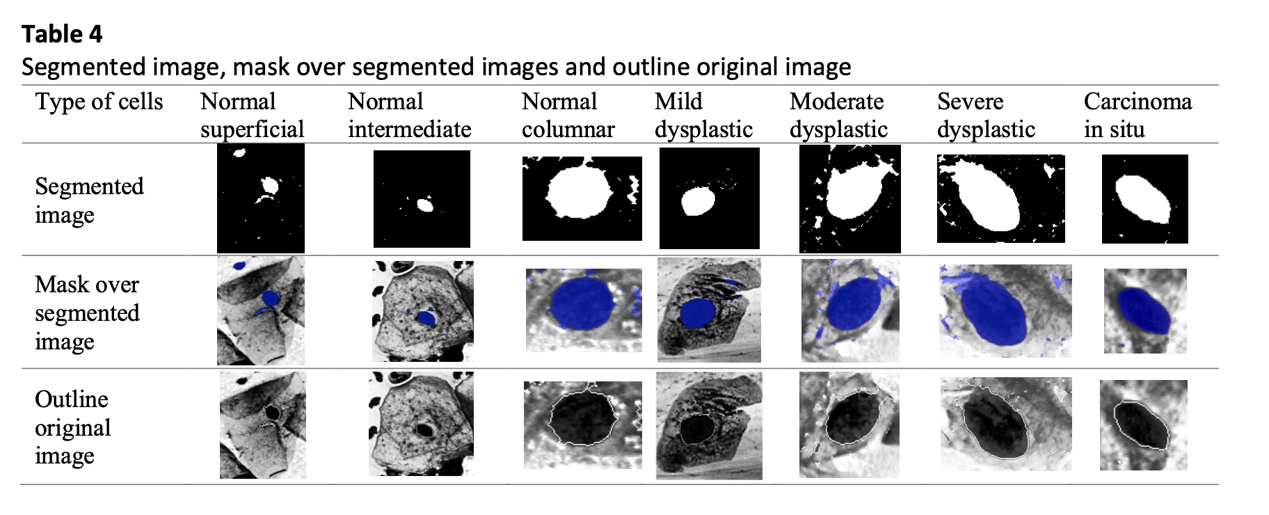
Cervical cancer is a prevalent and fatal disease that affects women all over the world. This affects roughly 0.5 million women annually and kills over 0.3 million people. Recently, a significant amount of literature has emerged around the advancement of technologies for identifying cervical cancer cells in women. Previously, diagnosing cervical cancer was done manually, which could lead to false positives or negatives. The best way of interpreting Pap smear images and automatically diagnose cervical cancer are still up for debate among the researchers. Method used in this study is the contrast enhancement technique for pre-processing and edge detection-based for segmentation of the nucleus. In this study, the average performance results of the method showed an accuracy of 96.99% in the seven-class problem using Herlev dataset. The present finding also support this study which concluded the results of accuracy achieved for the algorithm used for nucleus detection is improved by 6.15% when comparing to previous work. The accuracy value is in the lines of earlier literature that achieved accuracy of the approach used above 90% for seven class of cells. The major feature of the suggested approach is an improvement in the ability to anticipate which cells are aberrant and which are normal. Adding more classifiers could improve the suggested system even further. Therefore, a cervical cancer screening system might utilize this framework to identify women who have precancerous lesions.  

Downloads
Download data is not yet available.

Author Biographies

Nur Ain Alias, Faculty of Electrical Engineering Technology, Universiti Malaysia Perlis, UniCITI Alam Campus, Sungai Chuchuh, 02100 Padang Besar, Perlis, Malaysia

ainalias@studentmail.unimap.edu.my

Wan Azani Mustafa, Advanced Computing (AdvCOMP), Centre of Excellence, Universiti Malaysia Perlis (UniMAP), Pauh Putra Campus, 02600 Arau, Perlis, Malaysia

azani.mustafa@gmail.com

Mohd Aminuddin Jamlos, Faculty of Electronic Engineering Technology, Universiti Malaysia Perlis, UniCITI Alam Campus, Sungai Chuchuh, 02100 Padang Besar, Perlis, Malaysia

mohdaminudin@unimap.edu.my

Mohd Wafi Nasrudin, Faculty of Electronic Engineering Technology, Universiti Malaysia Perlis, UniCITI Alam Campus, Sungai Chuchuh, 02100 Padang Besar, Perlis, Malaysia

wafi@unimap.edu.my

Muhammad As'syarafi Mansor, Bagan Hospital Specialist Centre, Jalan Bagan Satu, 13400 Butterworth, Pulau Pinang, Malaysia

massyarafim@bagan.com.my

Hiam Alquran, Department of Biomedical Systems and Informatics Engineering, Yarmouk University 556, Irbid 21163, Jordan

heyam.q@yu.edu.jo

Published

2022-09-15

Issue

Section

Articles

Most read articles by the same author(s)

1 2 3 > >>