Detection of Kidney Stone and Estimation of its Size using Image Segmentation Techniques

Authors

  • Shraman Jain School of Electrical Engineering, Vellore Institute of Technology, Vellore, 632014 Tamil Nadu, India
  • Rajini G. K. School of Electrical Engineering, Vellore Institute of Technology, Vellore, 632014, Tamil Nadu, India
  • Rahul S.G. Department of Electronics and Communication Engineering, Vel Tech Rangarajan Dr. Sagunthala R&D Institute of Science and Technology, 600062 Chennai, India
  • Rajkumar R. Department of Electronics and Communication Engineering, Vel Tech Rangarajan Dr. Sagunthala R&D Institute of Science and Technology, 600062 Chennai, India
  • Velmurugan S. Department of Biomedical Engineering, Dr. N.G.P. Institute of Technology, 641048 Coimbatore, India
  • M. Jasmine Pemeena priyadarsini School of Electrical Engineering, Vellore Institute of Technology, Vellore, 632014, Tamil Nadu, India

DOI:

https://doi.org/10.37934/araset.36.2.91109

Keywords:

Kidney stones, Size, CT, Watershed segmentation

Abstract

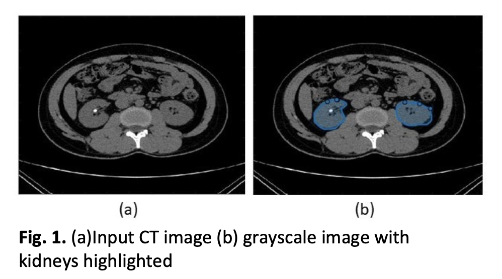
The study discusses the development of a reliable strategy for detecting and segmenting kidney stones from CT scans. Kidney stones are solid materials that can form in the kidneys due to excessive amounts of specific minerals in the urine. Severe back, side, lower abdominal, or groin discomfort, or blood in urine can indicate kidney stone presence. The current method for detection using CT scans is widely used, but it requires more precise and efficient technology due to the laborious and time-consuming manual process. The objective of this report is to propose a robust approach for kidney stone detection and segmentation, employing threshold segmentation and the watershed algorithm along with pre-and post-image processing for noise reduction. Additionally, the method estimates the size of the stone, aiding in selecting the appropriate medical procedure for stone removal. The entire process is implemented on a dataset comprising numerous kidney stone images to test and validate its accuracy and reproducibility. The system's image quality enhancement is evaluated by calculating several parameters such as SSIM, PSNR, FSIM, NCC, and NAE on the output images. To further validate the suggested method, a comparison is made with two other existing algorithms based on the mentioned evaluation criteria. The proposed strategy promises to improve the accuracy and reliability of kidney stone detection and segmentation, providing a more efficient and effective solution for medical diagnosis and treatment

Author Biographies

Shraman Jain, School of Electrical Engineering, Vellore Institute of Technology, Vellore, 632014 Tamil Nadu, India

shraman.jain2019@vitstudent.ac.in

Rajini G. K., School of Electrical Engineering, Vellore Institute of Technology, Vellore, 632014, Tamil Nadu, India

rajini.gk@vit.ac.in

Rahul S.G., Department of Electronics and Communication Engineering, Vel Tech Rangarajan Dr. Sagunthala R&D Institute of Science and Technology, 600062 Chennai, India

rahulgopi1993@yahoo.com

Rajkumar R., Department of Electronics and Communication Engineering, Vel Tech Rangarajan Dr. Sagunthala R&D Institute of Science and Technology, 600062 Chennai, India

rajkumarramasami@gmail.com

Velmurugan S., Department of Biomedical Engineering, Dr. N.G.P. Institute of Technology, 641048 Coimbatore, India

velmurugan.s@drngpit.ac.in

M. Jasmine Pemeena priyadarsini, School of Electrical Engineering, Vellore Institute of Technology, Vellore, 632014, Tamil Nadu, India

jasmin@vit.ac.in

Downloads

Published

2023-12-30

How to Cite

Shraman Jain, G. K., R., S.G., R. ., Rajkumar R., S., V., & M. Jasmine Pemeena priyadarsini. (2023). Detection of Kidney Stone and Estimation of its Size using Image Segmentation Techniques. Journal of Advanced Research in Applied Sciences and Engineering Technology, 36(2), 91–109. https://doi.org/10.37934/araset.36.2.91109

Issue

Section

Articles